Evolved EngineeringArchitecture
Potent CPU+GPU Core
Integrated with upgraded hardware platform, the processing capability and efficiency of S50 ELITE are boosted by several times*; therefore, users are given an enhancing scanning with accelerated imaging and exquisite clarity.
High-performance Transducers
Single Crystal Convex C1-6
Perfect uniformity of crystal alignment generates pure imaging with better penetration and higher S/N ratio, especially for difficult patients mainly in abdominal and OB applications.
Single Crystal Sector S1-5
Innovative single crystal material effectively enhances energy transmission and elevates spatial resolution for better clarity, mainly for cardiology and transcranial applications.
Composite Crystal Linear Transducers
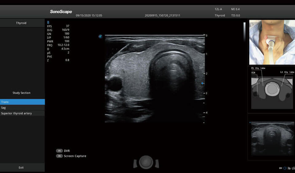
Better acoustic spectrum and lower acoustic impedance technology serve well in vascular, breast, thyroid, MSK, etc. The combo of 12L-A, 12L-B, 9L-A covers an ultra-wide frequency bandwidth, leaving nearly no blind spot for all sorts of scanning.
Ultra-light Crafted Volume VC2-9
Reduced weight, ultra-wide bandwidth, exquisite resolution and penetration at high volume rate, VC2-9 is a one-probe-solution throughout nearly the entire pregnancy.
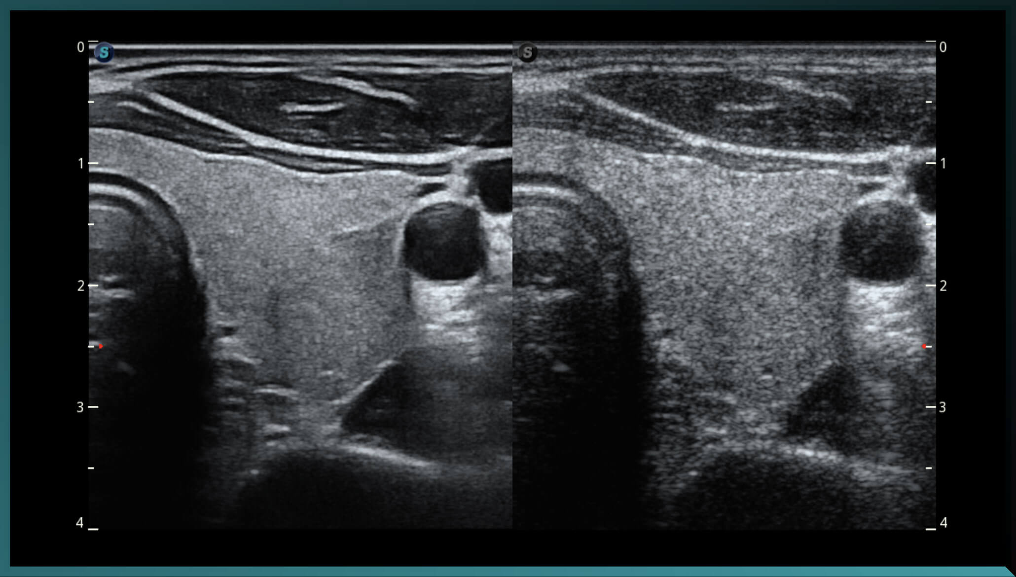
μScan+
Available for both B and 3D/4D modes, the new generation μScan+ provides you authentic presentation of details and lesion display through speckle reduction and enhanced border continuity.
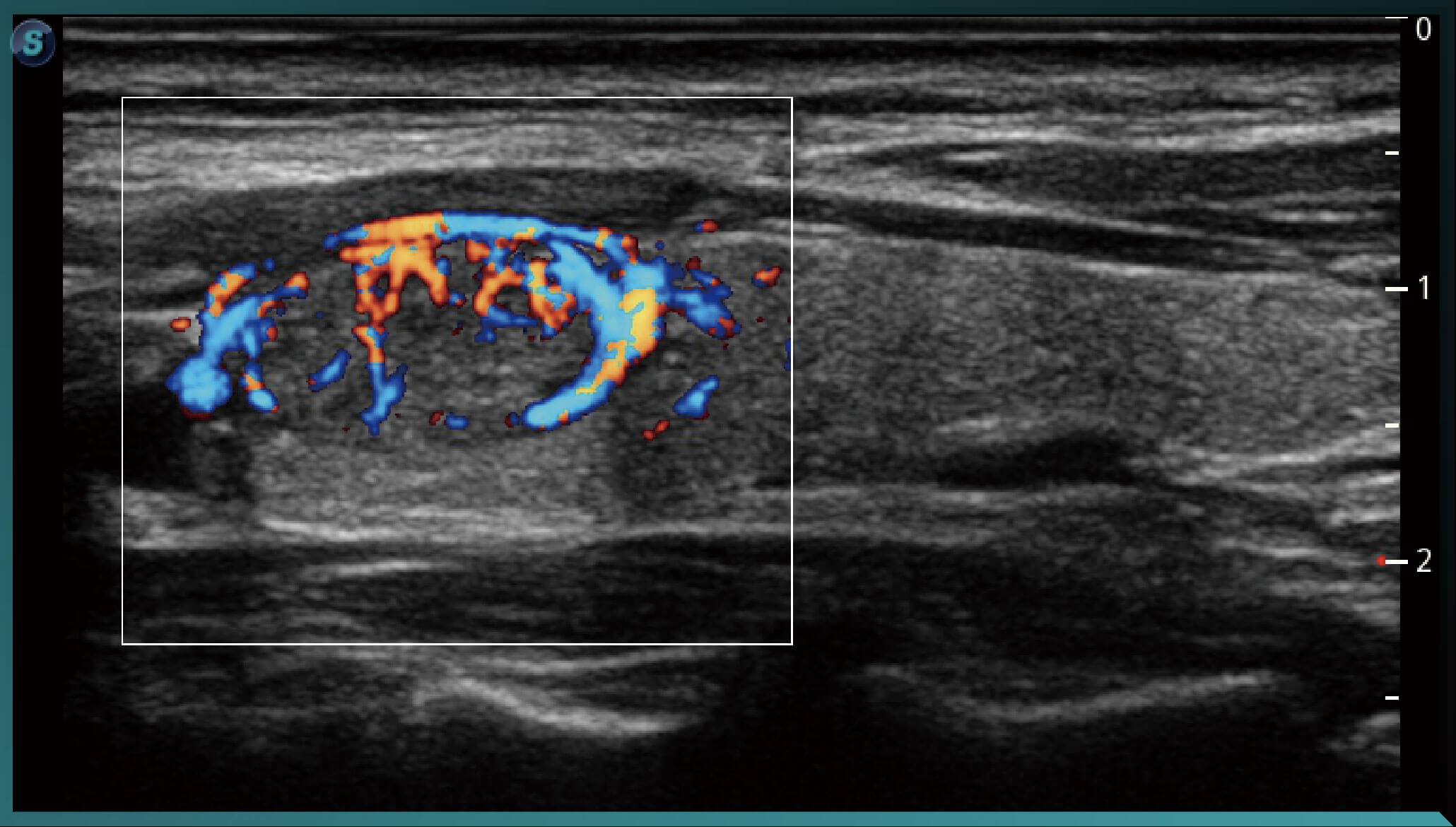
SR-Flow
Highly effective filter technology visualizes slow flows, enabling a vivid Doppler display with high sensitivity.
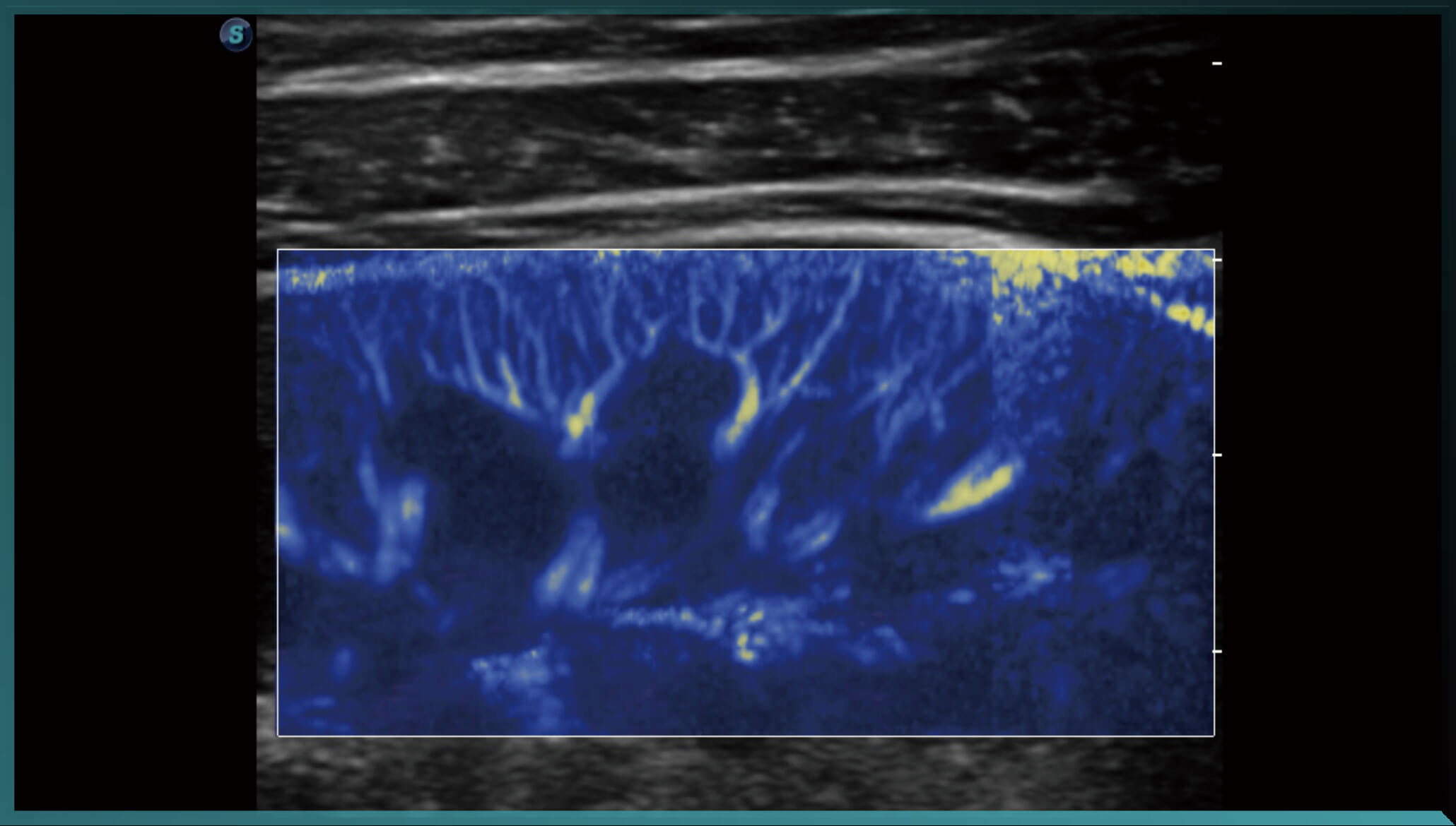
Micro F
Micro F provides an innovative method to expand the range of visible flow in ultrasound, especially for visualizing hemodynamic of tiny vessels.
Bright Flow
3D-like color Doppler flow strengthens boundary definition of vessel walls, without the need of using volume transducer.
Intelligent Solution
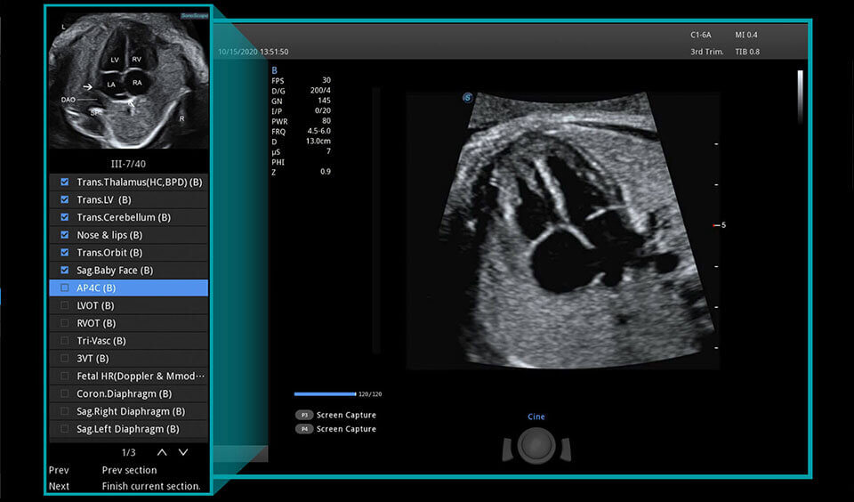
S-fetus
Realize automatic standard plane data collection and measurement using artificial intelligence technologies, with a single click. Common fetal biometric results are generated accurately and efficiently
Auto OB
A deep learning-based method that helps efficiently acquire fetal biometrics and reduces operator-dependent variability.
Auto NT
Provides semi-automated, standardized measurements of nuchal translucency on 2D images, reducing operator dependency..
Auto Bladder
Essential for bladder wall delineation and volume measurement, providing accurate contours and results regardless of bladder shape and size.
Automatikus EF
Auto EF calculates the ejection fraction based on LV wall tracing and Simpson’s rule. The simplified workflow saves time and effort.
Auto Face
Auto Face optimizes fetal 3D face visualization for easier abnormality diagnosis by removing obstructions and artifacts.
Auto B/C
Image parameter tuning no longer requires tedious effort. Auto B/C optimizes image quality in B and color Doppler modes with a single click
AVC Follicle
AVC Follicle provides high-efficiency follicle analysis through volume-based automatic calculation, including follicle count and volume.
Talented Features
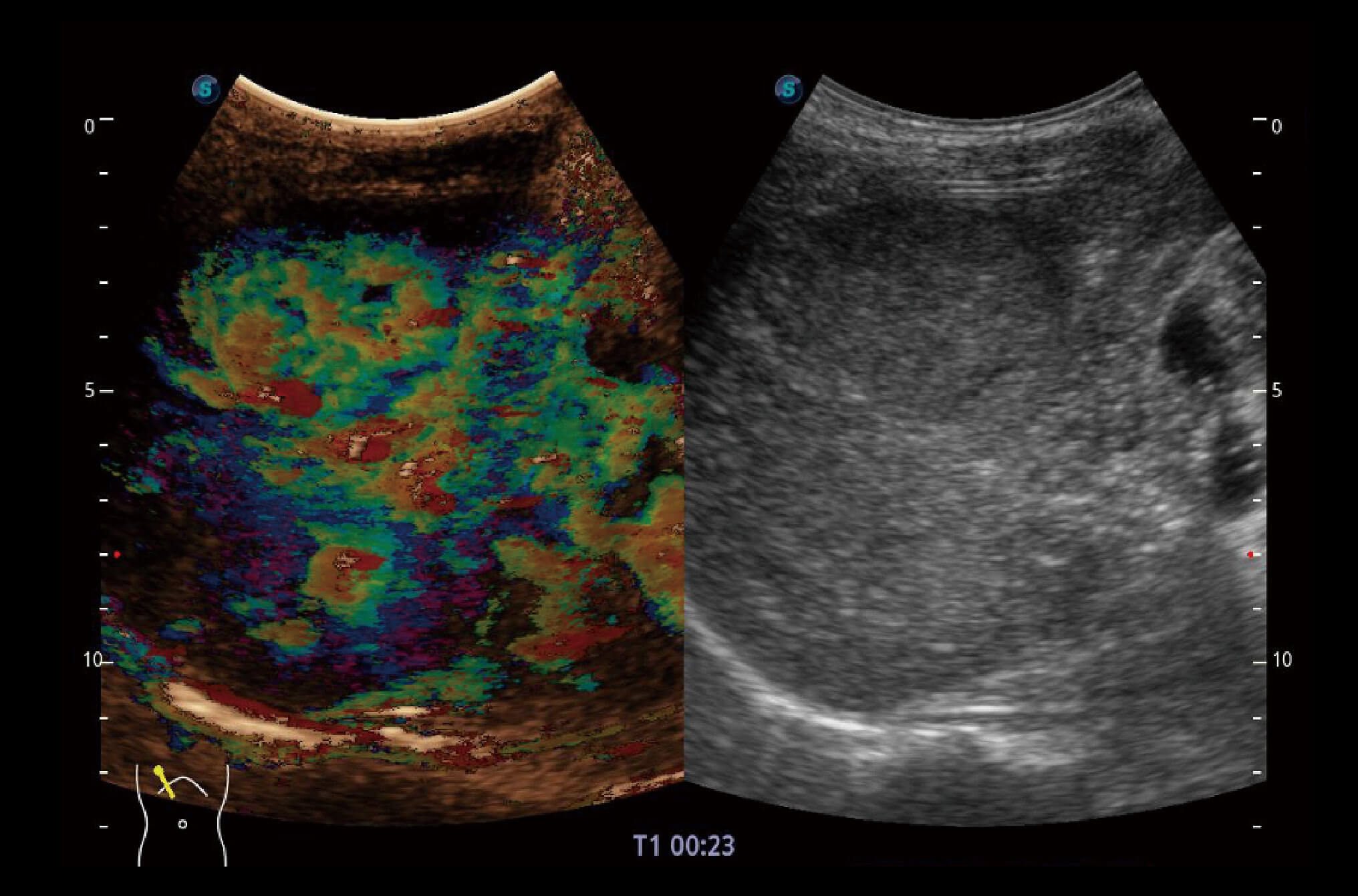
CEUS with MFI
Enhanced perfusion display traces small bubble populations, even in low-perfused and peripheral regions.
MFI-Time
Color-coded parametric view showing contrast uptake times during various perfusion phases for better tissue differentiation. Assists in assessing tumor blood flow distribution.
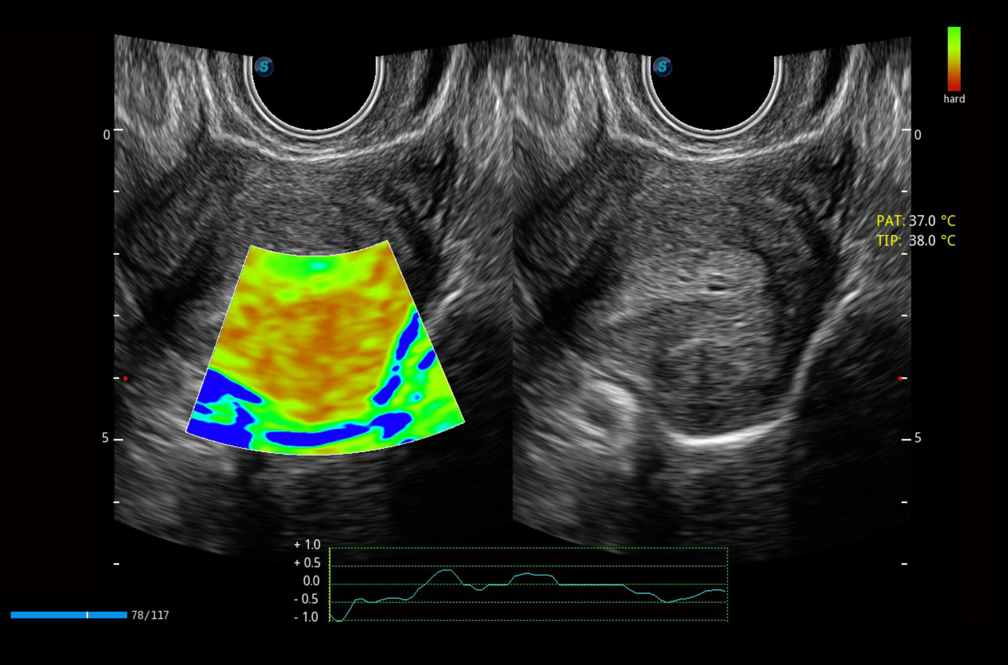
Strain Elastography
Real-time tissue stiffness assessment displayed via a color map to detect potential abnormalities in normal tissues. Available on linear, convex, and transvaginal probes, covering a wide range of regions. Semi-quantitative analysis based on strain ratio between lesion and normal tissue shows relative stiffness.
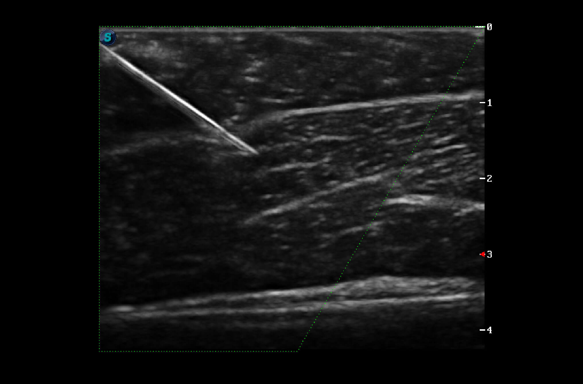
Vis-Needle
With beam steering, Vis-Needle improves visibility of the needle shaft and tip, enhancing diagnostic accuracy and enabling safe, precise procedures like nerve blocks.
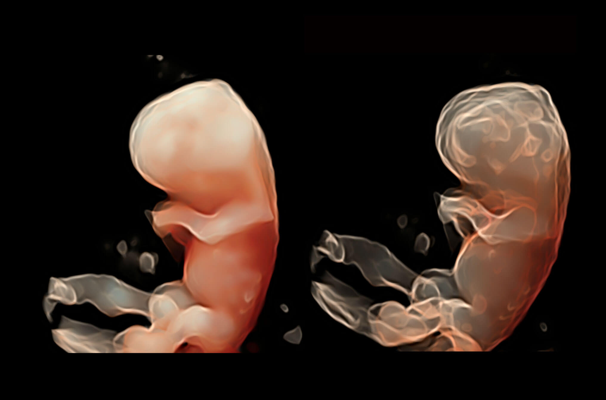
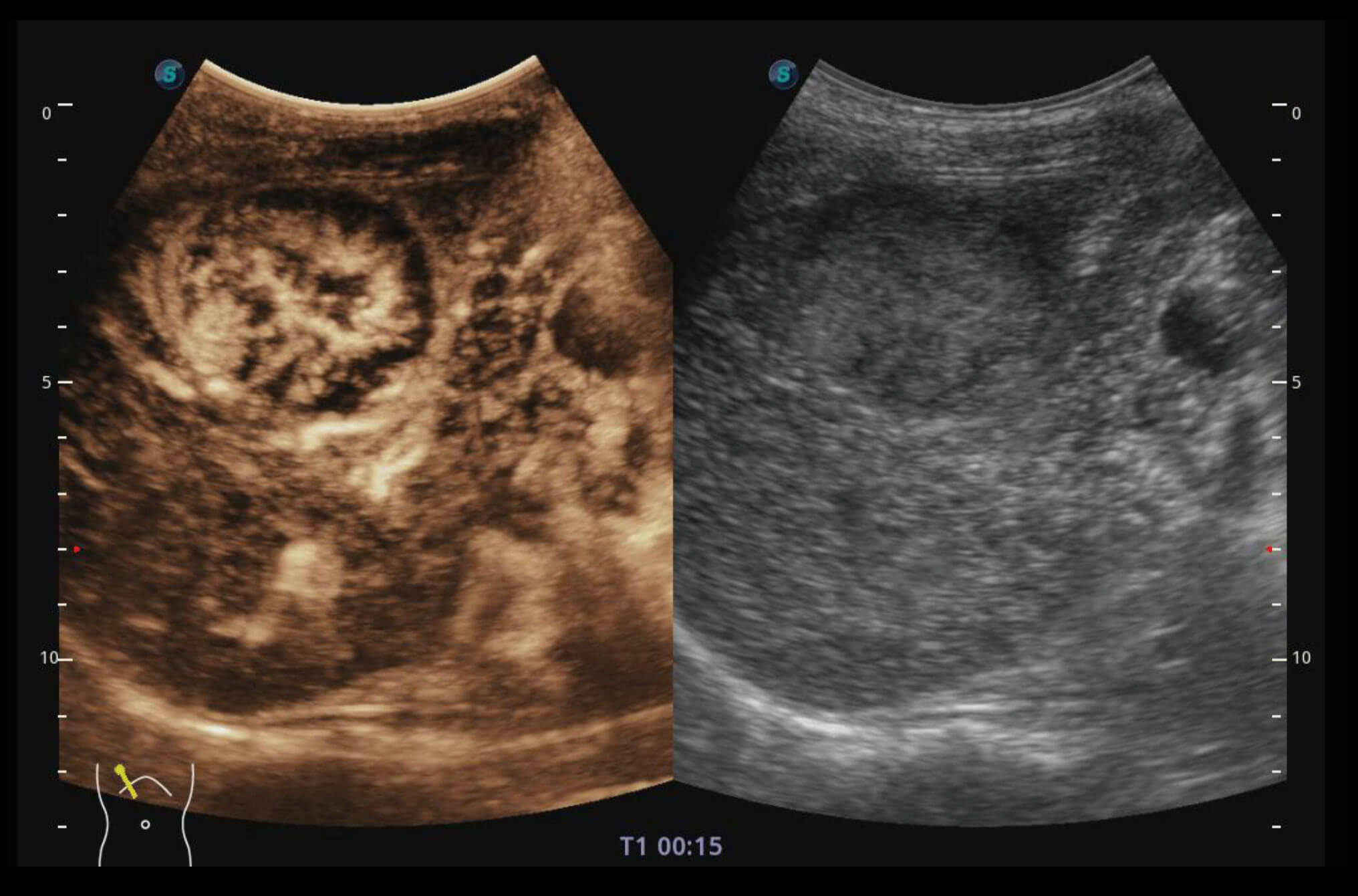
S-Live and S-Live Silhouette
S-Live enables detailed visualization of fine anatomical structures with a movable virtual light source, while S-Live Silhouette provides transparent volume images for comprehensive internal and external anatomical views—supporting intuitive diagnosis with real-time 3D imaging and improving patient communication.
Color 3D
Available in color and Power Doppler modes, Color 3D applies advanced rendering techniques—including S-Live and S-Live Silhouette—to display blood flow intuitively and naturally within vascular networks such as the umbilical cord.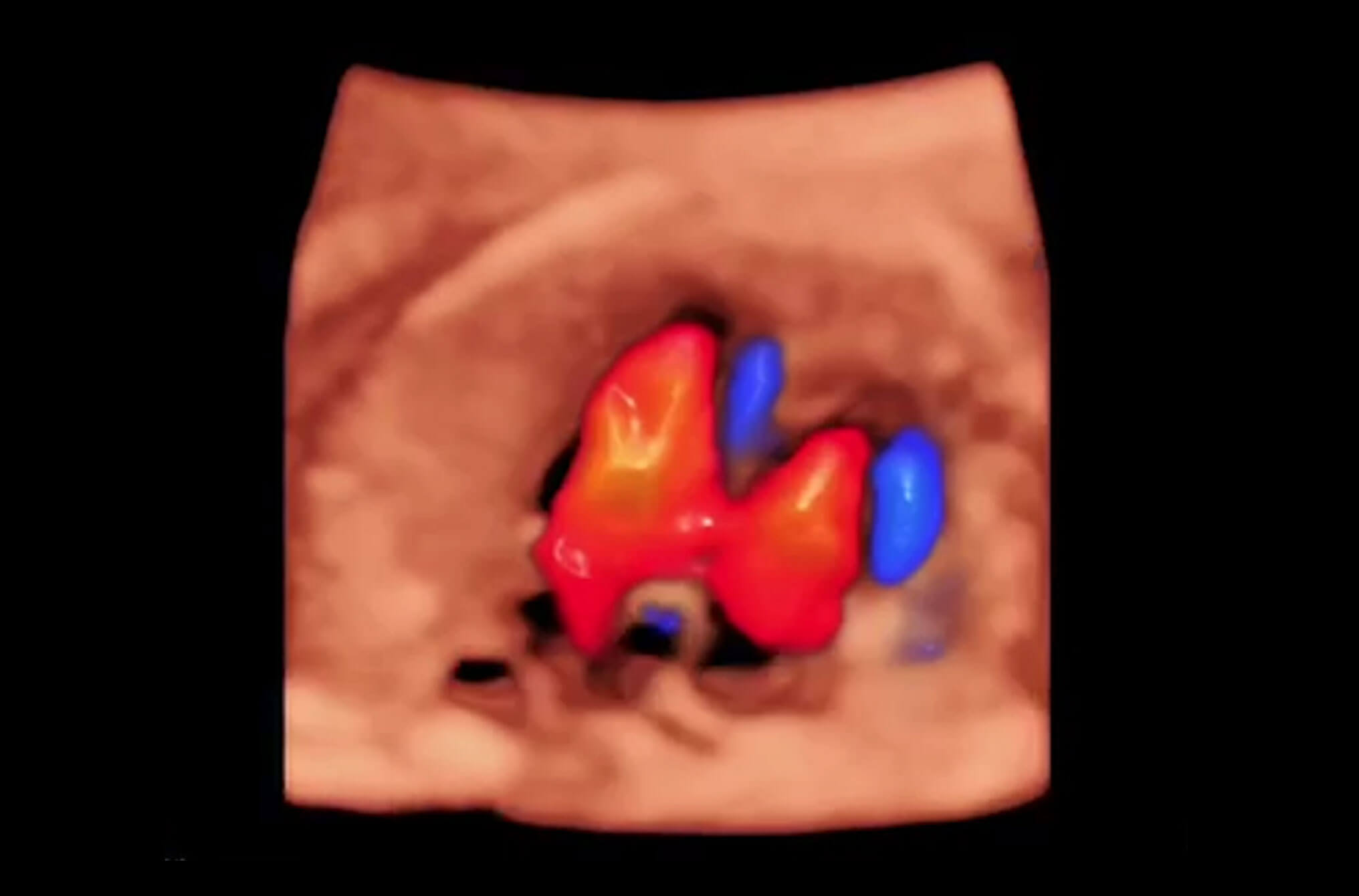
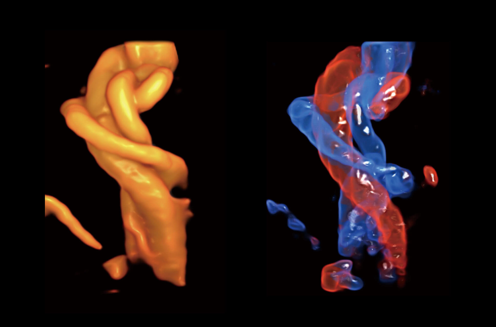
Spatio-Temporal Image Correlation (STIC)
Rapidly captures and visualizes fetal anatomical structures in motion, aiding the diagnosis of congenital heart disease.
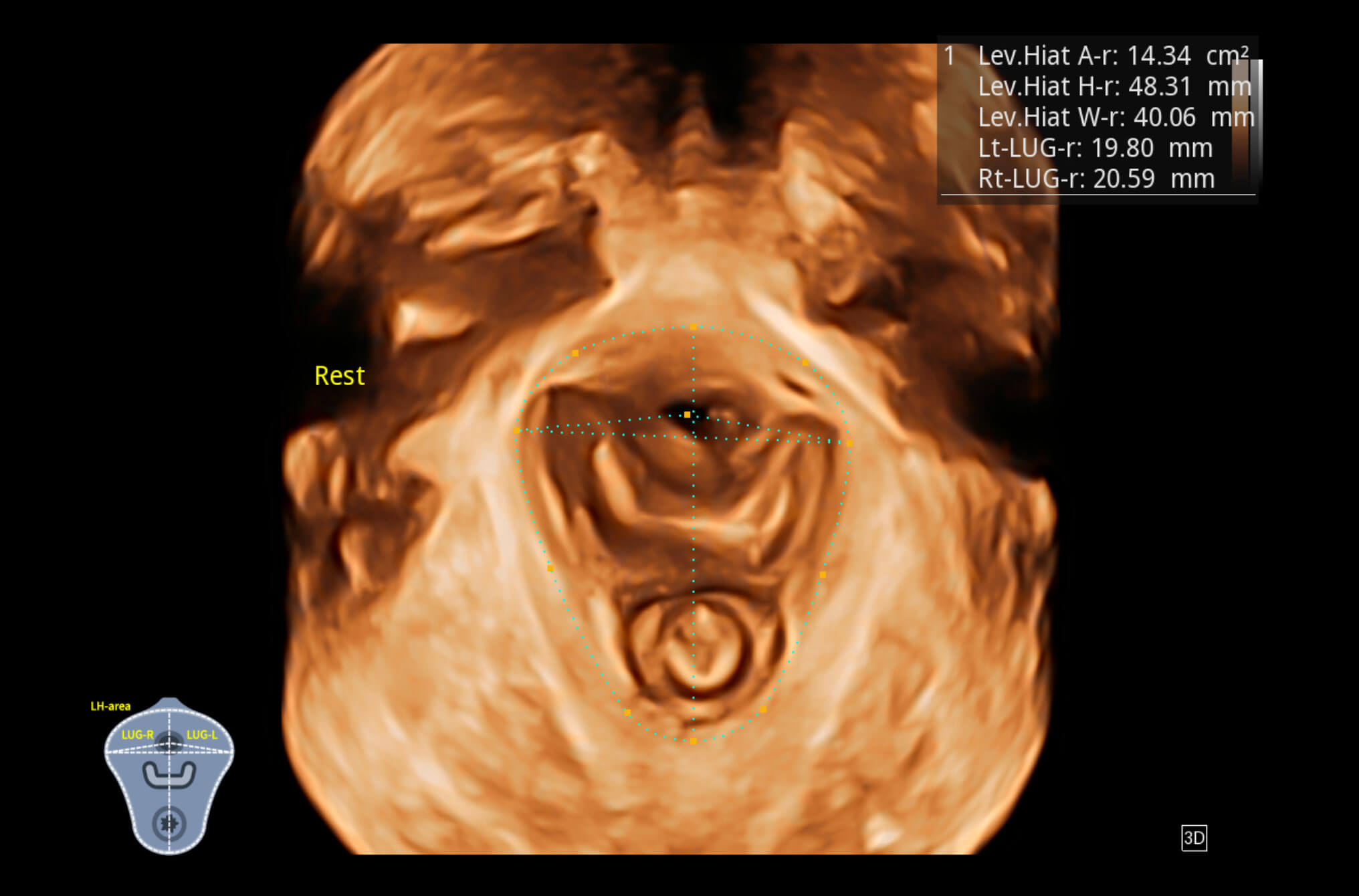
Pelvic Floor Imaging
Together with dedicated transvaginal probes, both 2D and volume imaging are available for detailed evaluation of pelvic anatomy including muscles, bladder, uterus, and more.
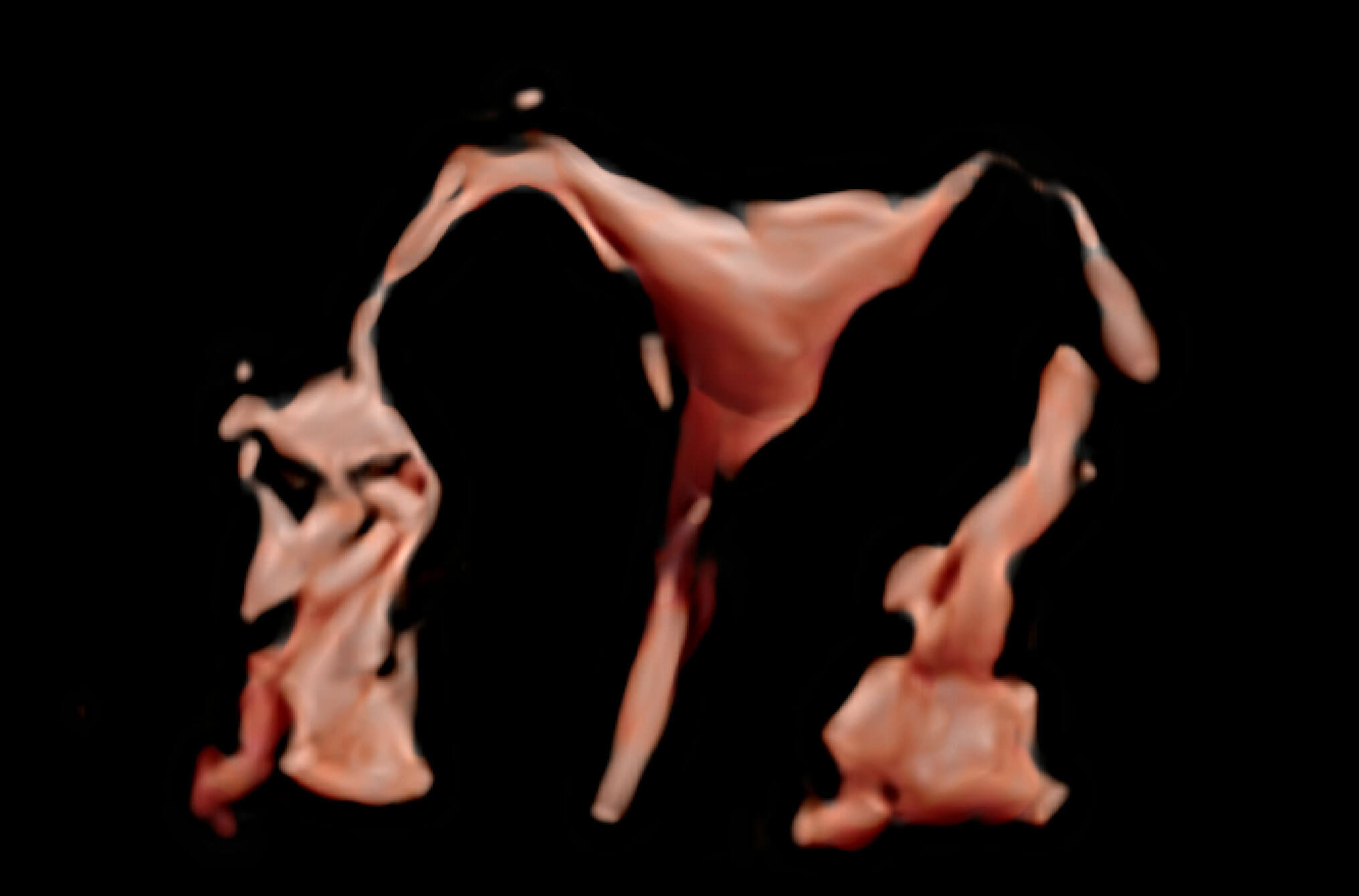
4D Hysterosalpingo-Contrast Sonography (HyCoSy)
Real-time 4D HyCoSy enables intuitive and confident gynecological evaluation of fallopian tube patency and improves 3D visualization of uterine and tubal morphology. Real-time perfusion process is clearly visible, providing additional information for accurate patency diagnosis.
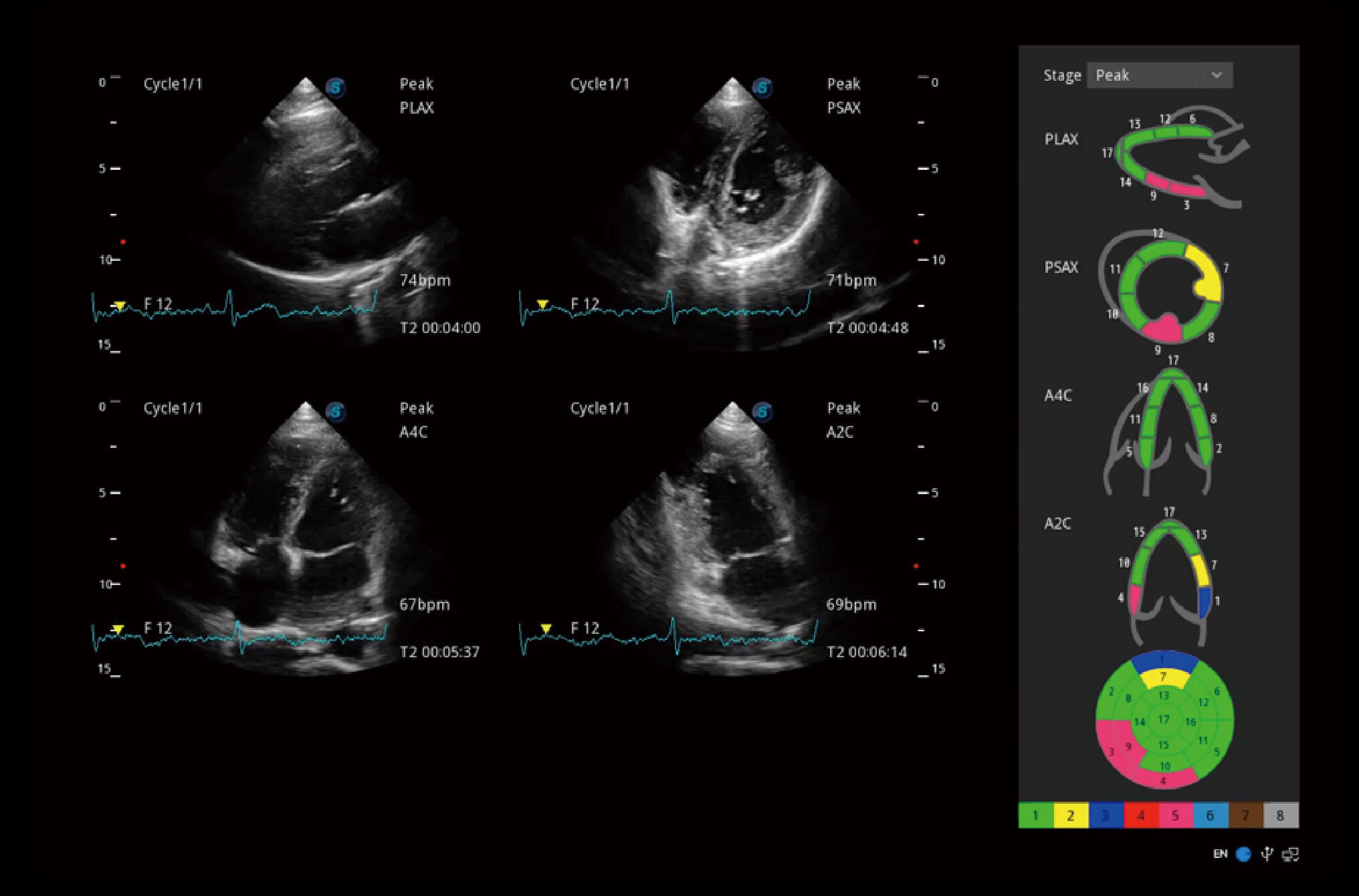
Stress Echo
A simple template for clinicians to perform stress echocardiography. Professional wall motion scoring and reporting provide visualized, intuitive results.
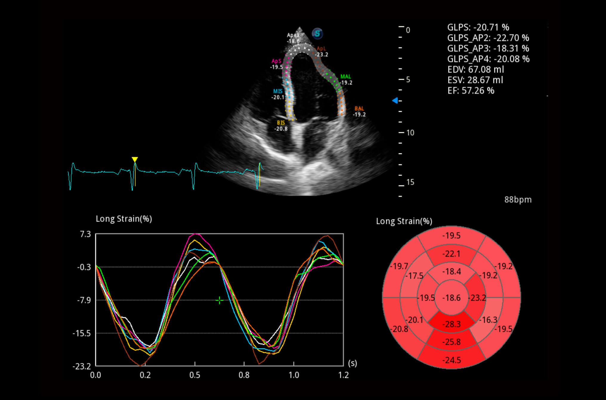
Myocardial Quantitative Analysis (MQA)
MQA provides precise quantitative measurement of myocardial mechanics using real-time, sensitive wall motion tracking. Offers global and regional assessments including strain, strain rate, displacement, velocity, etc.
LVO
Enhanced capabilities of the S50 Elite allow for LV opacification during stress exams, improving differentiation between myocardium and blood pool, and enhancing endocardial border visualization—especially in difficult patients.
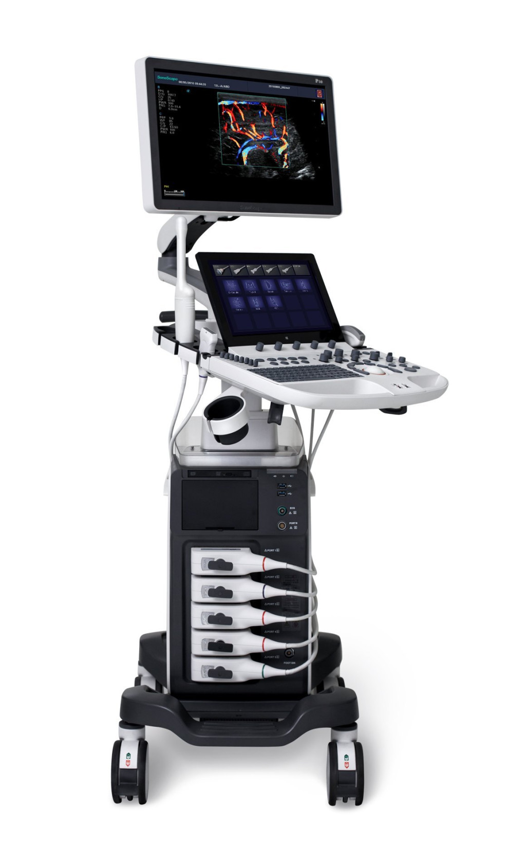
Pleasing Design
The design of S50 Elite took operational use into consideration, creating a comfortable diagnosing environment. Ergonomic design, excellent man-machine interaction and rapid response ensure S50 Elite a better user experience, bringing improved efficiency and helping to prevent fatigue from multiple examinations.
Sono-Help
Sono-Help is an inspiring tutorial displaying probe placement, anatomy illustration and standard ultrasound image examples. As a useful reference less experienced clinicians could rely on, Sono-Help covers a variety of applications including liver, kidney, cardiac, breast, thyroid, obstetrics, vascular, etc.
Sono-Assistant
Sono-assistant guides clinicians through the entire exam and provides customizable scanning protocol helps streamline workflow while increasing standardization and reducing keystrokes and exam time.

Sono-Drop
Sono-drop provides a fast and convenient ultrasound image transmission between S50 Elite and the patients’ smart devices. The bond between clinicians and patients are supposed to be strengthened through more frequent communication.

Sono-Synch
Real-time interface and camera sharing, enabled by Sono-synch, makes it possible to connect two ultrasound in a remote distance and perform remote medical consultation and tutorial.