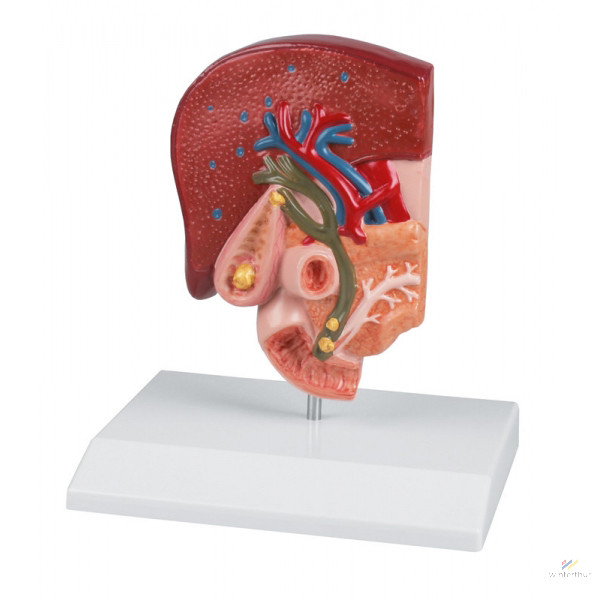
This semi-life-size model shows in detail the anatomy and environment of the bile ducts. The gallbladder wall shows tissue lesions caused by both chronic inflammation and acute inflammation (cholecystitis). Gallstones can be seen in the following typical locations:
- spiral valve- gallbladder bed- common bile duct- papillary opening into the small intestine.
Mounted on a base.
Product weight: 0,5 kg
